Anti-human TJP2 / ZO-2 Recombinant Monoclonal Antibody (HM0055)
Price range: $180.00 through $480.00
Anti-human ZO-2 Recombinant Monoclonal Antibody (HM0055) is a high-affinity recombinant monoclonal antibody generated by phage-display selection from a mouse-derived antibody library followed by structure-guided affinity optimization. The antibody was validated by ELISA against recombinant epitope-Fc fusion antigen, by Western blot against recombinant MBP-epitope fusion protein, and by Western blot detection of endogenous full-length ZO-2 protein.
Also known as: hZO-2, ZO-2, ZO2, TJP2, and Tight junction protein 2.
For research use only. Not for diagnostic or therapeutic applications.
Product Information
| Property | Description |
|---|---|
| Product Name | Anti-human TJP2 / ZO-2 Recombinant Monoclonal Antibody |
| Antibody ID | HM0055 |
| Target Protein | Tight junction protein 2 |
| Target Protein Short Name | ZO-2 |
| UniProt Accession | Q9UDY2-1, Q9UDY2-2, Q9UDY2-3, Q9UDY2-4, Q9UDY2-5, Q9UDY2-6, Q9UDY2-7 |
| Molecular Weight | 111.7–137.3 kDa depending on isoform |
| Antibody Type | Recombinant monoclonal antibody |
| Host Species | Mouse |
| Recombinant Format | Full-length mouse IgG1 |
| Isotype | Mouse IgG1 |
| Species Reactivity | Human |
| Tested Applications | WB |
| Recommended Dilution for WB | 1:500–1:5000 |
| Antigen | Recombinant epitope-Fc fusion protein |
| Epitope Sequence | DSDSEIEDISEIESNRSFSPEE |
| Expression System | HEK293 cells |
| Purification | Protein G affinity chromatography |
| Conjugate | Unconjugated |
| Concentration | 0.1 mg/mL |
| Storage Buffer | 0.1 M Tris, 0.05 M Glycine, 0.07 M NaCl, 2 g/L BSA, 50% glycerol, pH 7 |
| Storage Condition | Store at -20°C |
| Research Use | For research use only. Not for diagnostic or therapeutic applications |
Product Background
Tight junction protein 2 (ZO-2, Zona occludens-2) is a scaffold protein associated with epithelial and endothelial tight junctions. ZO-2 contributes to maintenance of cell polarity, epithelial barrier integrity, cytoskeletal organization, signal transduction, and regulation of intercellular junction complexes.
ZO-2 is widely studied in epithelial biology, barrier function, cancer metastasis, inflammation, gastrointestinal research, blood-brain barrier biology, and cell-cell junction signaling.
Antibody Development
HM0055 was generated by phage-display selection using a mouse-derived antibody library. Recombinant epitope-Fc fusion protein containing the ZO-2 epitope sequence was used as the selection antigen. Following identification of the initial antibody candidate, structure-guided affinity optimization was performed to improve binding affinity and specificity.
The optimized antibody was reconstructed as a full-length mouse IgG1 recombinant antibody and expressed in HEK293 cells.
Binding and specificity were evaluated by:
- ELISA using recombinant epitope-Fc fusion antigen
- Western blot detection of endogenous full-length ZO-2 protein
Validation Data
ELISA Validation
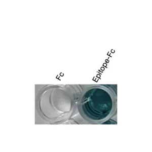
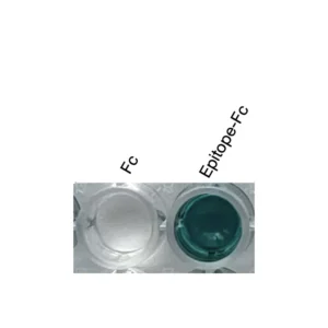
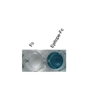
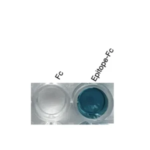
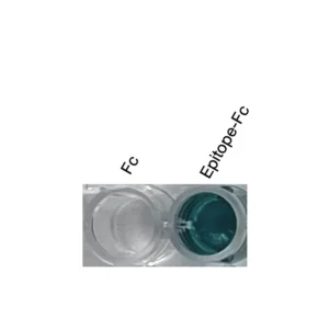
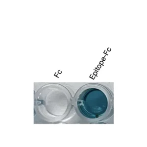
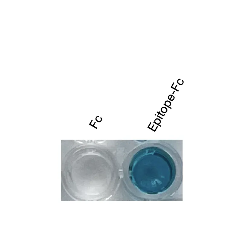
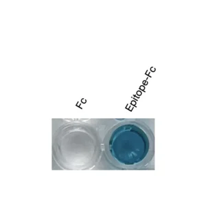
ELISA demonstrated specific binding to recombinant epitope-Fc fusion antigen, while minimal binding was observed against Fc-only control protein.
Validation conditions:
- Well 1: Fc control protein
- Well 2: Epitope-Fc fusion protein
- Detection substrate: TMB
Note:
ELISA validation confirms binding to epitope-Fc fusion antigen. Full-length protein binding in solution has not been tested by ELISA.
Western Blot Validation – Recombinant Fusion Protein
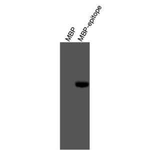
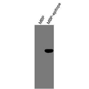
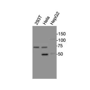
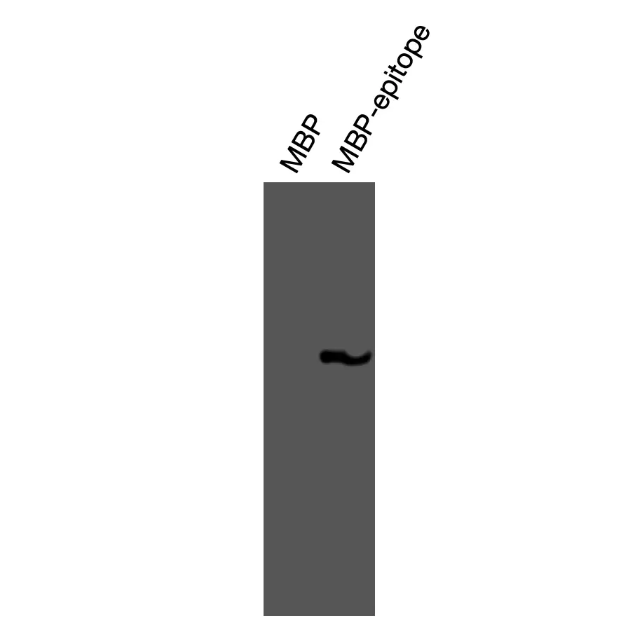
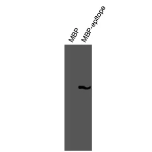
Western blot analysis demonstrated specific recognition of recombinant MBP-epitope fusion protein expressed in BL21(DE3) cells, while minimal binding was observed against MBP control protein.
Validation conditions:
- Lane 1: MBP control protein
- Lane 2: MBP-epitope fusion protein
- Standard reducing Western blot
Western Blot Validation – Endogenous Full-Length Protein
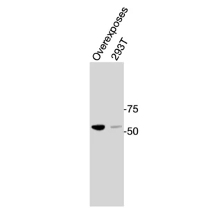
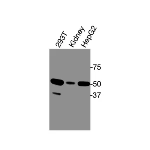
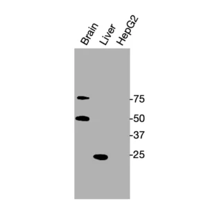
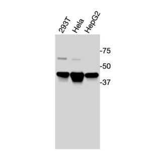
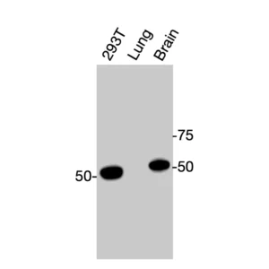
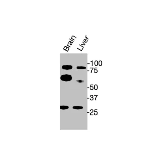
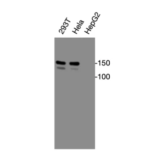
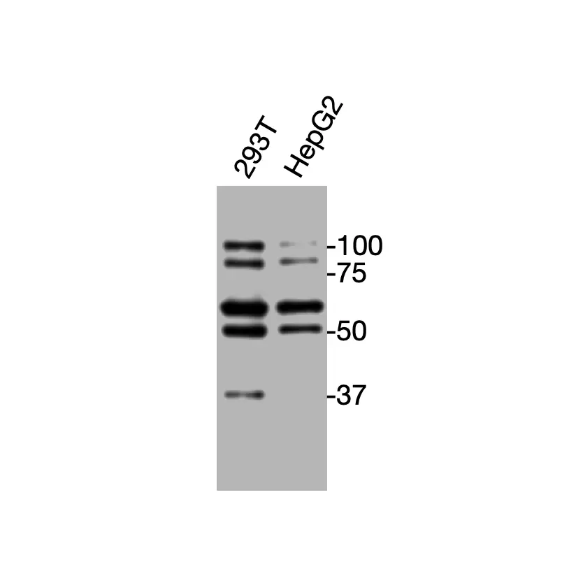
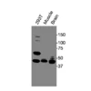
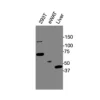
Western blot analysis detected endogenous full-length ZO-2 protein under reducing conditions.
Validated samples:
- Human HEK293T (293T) cells
- Human HepG2 cells
Observed bands:
- Multiple immunoreactive bands were detected.
- Lower molecular weight bands may represent cleavage products, alternatively processed isoforms.
Detection condition:
- Standard reducing Western blot
- HRP-conjugated anti-mouse secondary antibody
| Size | 20 ul, 100 ul |
|---|
Related products