
Anti-mouse/human GR Recombinant Monoclonal Antibody (XM0009)
Price range: $180.00 through $480.00
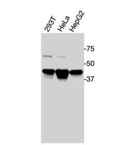
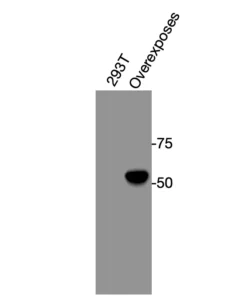
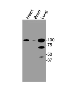
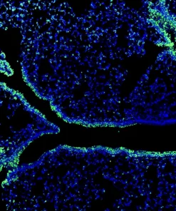
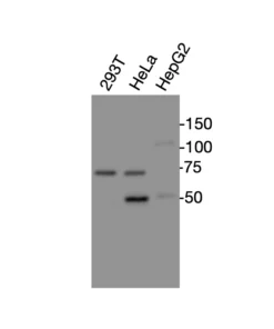
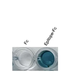
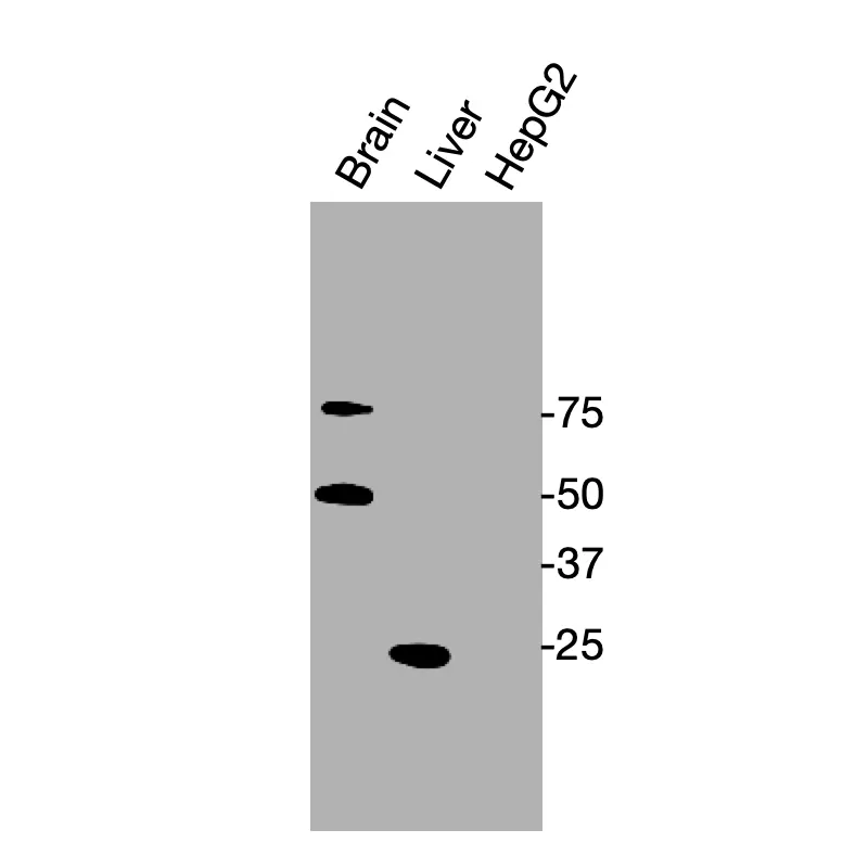
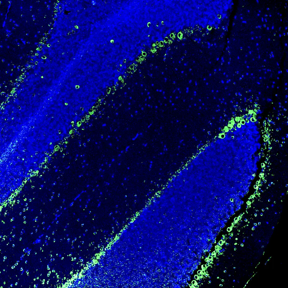
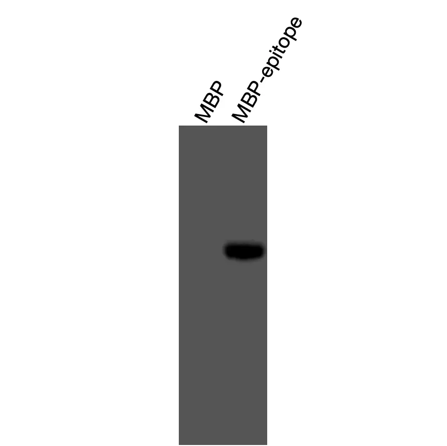
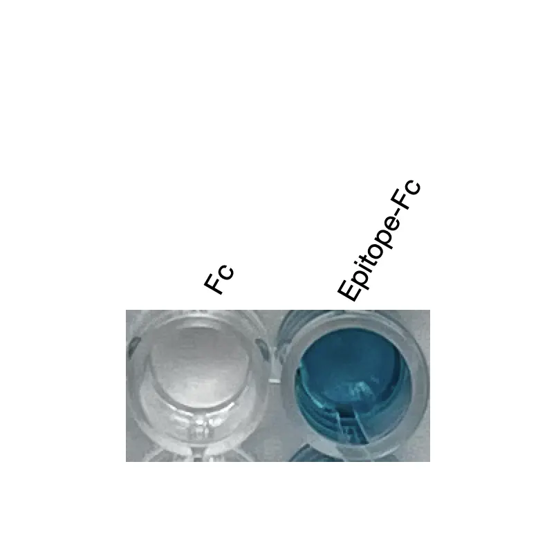
Anti-mouse/human GR Recombinant Monoclonal Antibody (XM0009) is a high-affinity recombinant monoclonal antibody generated by phage-display selection from a mouse-derived antibody library, followed by structure-guided affinity optimization. The antibody recognizes both mouse and human GR through a conserved epitope (AA 31–48) located within the N-terminal flexible region of the glucocorticoid receptor (GR). It has been validated by ELISA using a recombinant epitope-Fc fusion antigen, by Western blot against a recombinant MBP-epitope fusion protein, by Western blot detection of endogenous GR protein in HepG2 cell lysates and mouse tissue lysates, and by immunofluorescence staining of mouse brain tissue.
Also known as: GR, NR3C1, Glucocorticoid receptor, and Glucocorticoid receptor alpha.
For research use only. Not for diagnostic or therapeutic applications.
Product Information
| Property | Description |
| Product Name | Anti-mouse/human GR Recombinant Monoclonal Antibody |
| Antibody ID | XM0009 |
| Target Protein | Glucocorticoid receptor (GR) |
| UniProt Accession | Human: P04150; Mouse: P06537 |
| Molecular Weight | Human isoforms: 60.6–85.8 kDa; Mouse isoforms: 83.0–87.3 kDa |
| Antibody Type | Recombinant monoclonal antibody |
| Host Species | Mouse |
| Recombinant Format | Full-length mouse IgG1 |
| Isotype | Mouse IgG1 |
| Species Reactivity | Mouse and human |
| Tested Applications | WB, IF, ELISA |
| Recommended Dilution for WB | 1:500–1:5000 |
| Antigen | Recombinant epitope-Fc fusion protein |
| Epitope Sequence | YKTLRGGATVKVSASSP |
| Expression System | HEK293 cells |
| Purification | Protein G affinity chromatography |
| Conjugate | Unconjugated |
| Concentration | 0.1 mg/mL |
| Storage Buffer | 0.1 M Tris, 0.05 M Glycine, 0.07 M NaCl, 2 g/L BSA, 50% glycerol, pH 7 |
| Storage Condition | Store at -20°C |
| Research Use | For research use only. Not for diagnostic or therapeutic applications. |
Product Background
Glucocorticoid receptor (GR) is a ligand-regulated nuclear receptor that mediates the biological effects of glucocorticoids. GR is involved in stress responses, immune regulation, inflammation, metabolism, development, and transcriptional control. Because glucocorticoid signaling is central to immunology, endocrinology, neuroscience, and pharmacology, GR is widely studied in disease biology and therapeutic research.
Antibody Development
XM0009 was generated by phage-display selection using a mouse-derived antibody library. Recombinant epitope-Fc fusion protein containing the GR epitope sequence was used as the selection antigen. Following identification of the initial antibody candidate, structure-guided affinity optimization was performed to improve binding affinity and specificity.
The optimized antibody was reconstructed as a full-length mouse IgG1 recombinant antibody and expressed in HEK293 cells.
Binding and specificity were evaluated by:
- ELISA using recombinant epitope-Fc fusion antigen
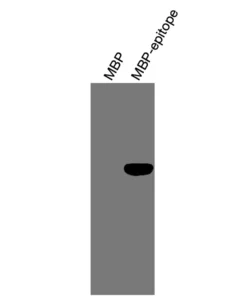
- Western blot using recombinant MBP-epitope fusion protein
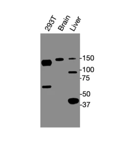
- Western blot detection of endogenous GR protein in mouse tissues and a human cell line
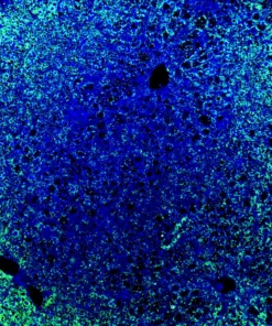
- Immunofluorescence staining of endogenous mouse GR in brain tissue
Validation Data
ELISA Validation
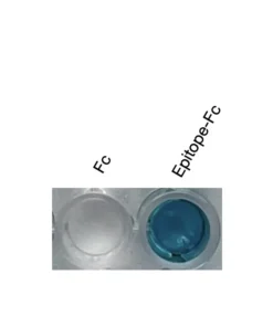
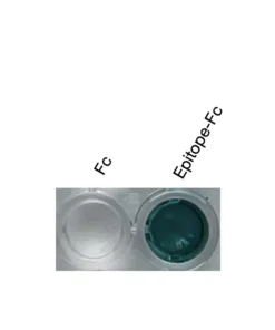
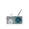
ELISA demonstrated specific binding to recombinant epitope-Fc fusion antigen, while minimal binding was observed against Fc control protein.
Validation conditions:
- Well 1: Fc control protein
- Well 2: epitope-Fc fusion protein
- Detection substrate: TMB
Note: ELISA validation confirms binding to the epitope-Fc fusion antigen. Full-length GR protein binding in solution has not been tested by ELISA.
Western Blot Validation – Recombinant Fusion Protein
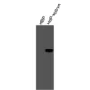
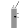
Western blot analysis demonstrated specific recognition of recombinant MBP-epitope fusion protein expressed in BL21(DE3) cells, while no significant binding was observed against MBP control protein.
Validation conditions:
- Lane 1: MBP control protein
- Lane 2: MBP-epitope fusion protein
- Standard reducing Western blot
Western Blot Validation – Endogenous Full-Length Protein
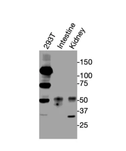
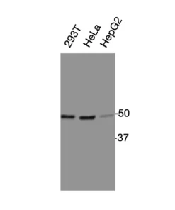
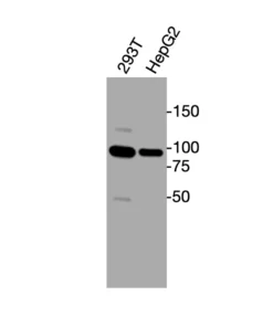
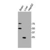
Western blot analysis detected endogenous GR protein in mouse tissues and a human cell line under reducing conditions.
Validated samples:
- Mouse brain tissue
- Mouse liver tissue
- HepG2 cells
Detection condition:
- Standard reducing Western blot
- HRP-conjugated anti-mouse secondary antibody
Immunofluorescence Validation
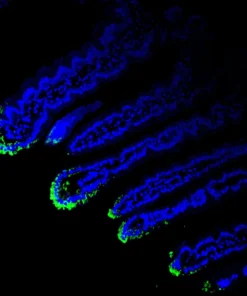
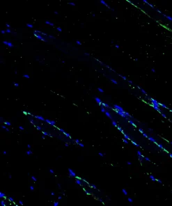
Immunofluorescence staining of mouse brain tissue showed endogenous GR-associated signal using XM0009. GR signal is shown in green and nuclei are counterstained with DAPI (blue).
| Size | 20 ul, 100 ul |
|---|
Related products