Anti-mouse/human PNR Recombinant Monoclonal Antibody (XM0028)
Price range: $180.00 through $480.00
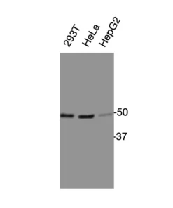
Anti-mouse/human PNR Recombinant Monoclonal Antibody (XM0028) is a high-affinity recombinant monoclonal antibody generated by phage-display selection from a mouse-derived antibody library, followed by structure-guided affinity optimization. The antibody recognizes both mouse and human PNR through a conserved epitope (AA 19-34) located within the N-terminal flexible region of photoreceptor-specific nuclear receptor (PNR). It has been validated by ELISA using a recombinant epitope-Fc fusion antigen, by Western blot against a recombinant MBP-epitope fusion protein, by Western blot detection of endogenous PNR protein in mouse tissue lysates, and by immunofluorescence staining of mouse lung tissue.
Also known as: Photoreceptor-specific Nuclear Receptor, NR2E3, PNR, Nuclear Receptor Subfamily 2 Group E Member 3.
For research use only. Not for diagnostic or therapeutic applications.
Product Information
| Property | Description |
|---|---|
| Product Name | Anti-mouse/human PNR Recombinant Monoclonal Antibody |
| Antibody ID | XM0028 |
| Target Protein | Photoreceptor-specific Nuclear Receptor |
| Target Protein Short Name | PNR (NR2E3) |
| UniProt Accession | Human: Q9Y5X4-1, Q9Y5X4-2; Mouse: Q9QXZ7 |
| Molecular Weight | Human isoform 1: 44.7 kDa; Human isoform 2: 39.6 kDa; Mouse: 43.2 kDa |
| Antibody Type | Recombinant Monoclonal Antibody |
| Host Species | Mouse |
| Recombinant Format | Full-length mouse IgG1 |
| Isotype | Mouse IgG1 |
| Species Reactivity | Mouse, Human |
| Tested Applications | WB, IF |
| Recommended Dilution for WB | 1:500–1:5000 |
| Recommended Dilution for IF | Optimize experimentally |
| Antigen | Recombinant epitope-Fc fusion protein |
| Epitope Sequence | KESPGRWGLGEDPTGV |
| Expression System | HEK293 cells |
| Purification | Protein G affinity chromatography |
| Conjugate | Unconjugated |
| Concentration | 0.1 mg/mL |
| Storage Buffer | 0.1 M Tris, 0.05 M Glycine, 0.07 M NaCl, 2 g/L BSA, 50% glycerol, pH 7 |
| Storage Condition | Store at -20°C |
| Research Use | For research use only. Not for diagnostic or therapeutic applications. |
Product Background
Photoreceptor-specific nuclear receptor (PNR, also known as NR2E3) is an orphan member of the nuclear receptor superfamily that functions as a ligand-dependent transcription factor regulating photoreceptor differentiation, retinal development, photoreceptor maintenance, and visual function. PNR plays an essential role in determining rod photoreceptor cell fate while suppressing cone-specific gene expression, thereby maintaining normal retinal homeostasis. Mutations in NR2E3 have been associated with several inherited retinal disorders, including enhanced S-cone syndrome (ESCS), retinitis pigmentosa, Goldmann-Favre syndrome, and other degenerative retinal diseases.
Beyond retinal development, increasing evidence suggests that NR2E3 contributes to transcriptional regulation, chromatin remodeling, cellular differentiation, and disease-associated signaling pathways. PNR has therefore become an important target for researchers investigating retinal biology, photoreceptor development, inherited eye diseases, nuclear receptor signaling, gene regulation, and transcription factor biology.
Antibody Development
XM0028 was generated by phage-display selection using a mouse-derived recombinant antibody library. A recombinant epitope-Fc fusion protein containing the PNR epitope sequence was used as the selection antigen. Following identification of the initial antibody candidate, structure-guided affinity optimization was performed to improve antigen-binding affinity while maintaining high specificity.
The optimized antibody was reconstructed as a full-length mouse IgG1 recombinant monoclonal antibody and expressed in HEK293 cells.
Binding specificity and performance were evaluated by:
- ELISA using recombinant epitope-Fc fusion antigen
- Western blot using recombinant MBP-epitope fusion protein
- Western blot detection of endogenous PNR protein in mouse tissues
- Immunofluorescence staining of mouse lung tissue
Validation Data
ELISA Validation
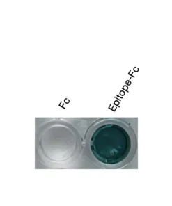
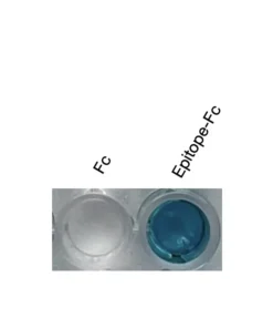
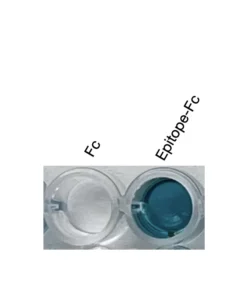
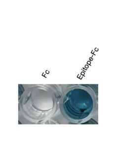
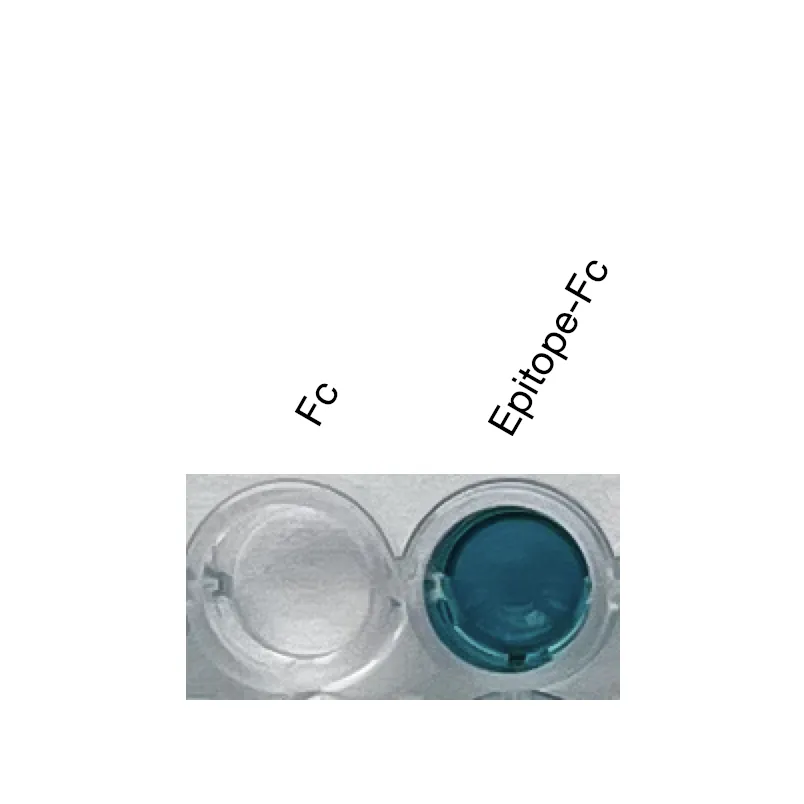
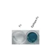
ELISA demonstrated strong and specific binding of XM0028 to the recombinant PNR epitope-Fc fusion protein, while minimal binding was observed against the Fc control protein. These results confirm that the antibody specifically recognizes the PNR epitope rather than the Fc fusion partner.
Validation conditions
- Fc control protein
- Recombinant PNR epitope-Fc fusion protein
- Detection substrate: TMB
Note: ELISA validation confirms binding to the recombinant peptide-Fc fusion antigen. Binding to full-length native PNR protein has not been evaluated by ELISA.
Western Blot Validation – Recombinant Fusion Protein
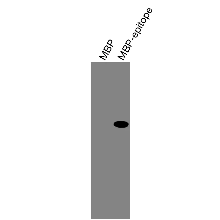
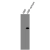
Western blot analysis demonstrated specific recognition of the recombinant MBP-PNR epitope fusion protein. No detectable signal was observed against the MBP control protein, confirming excellent specificity toward the designed PNR epitope.
Validation conditions
- MBP control protein
- Recombinant MBP-PNR epitope fusion protein
- Standard reducing Western blot
Western Blot Validation – Endogenous Full-Length Protein
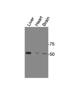
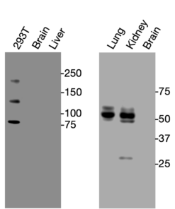
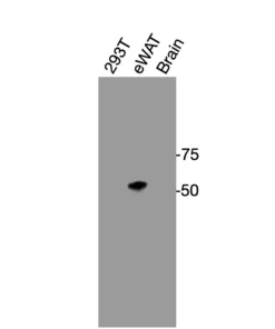
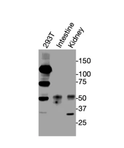
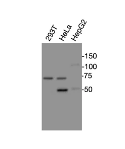
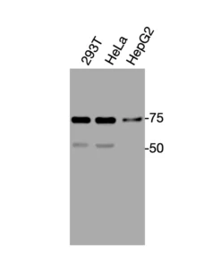
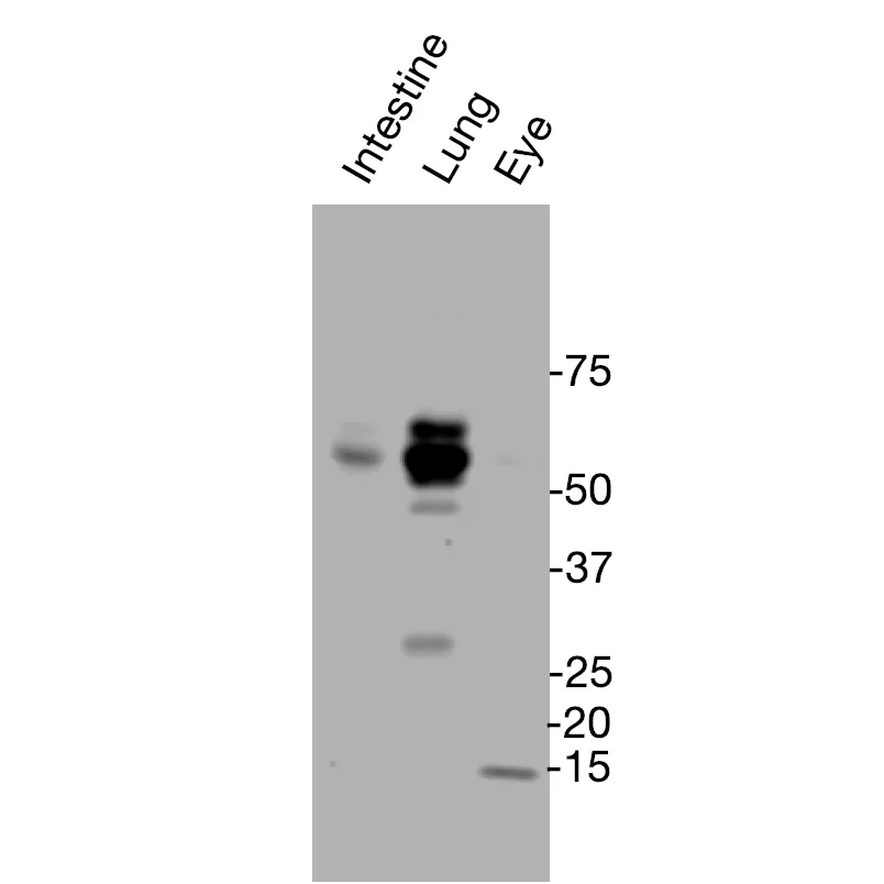
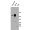
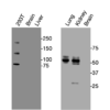
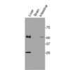
Western blot analysis detected endogenous PNR protein in mouse tissue lysates under reducing conditions.
Validated samples
- Mouse intestine
- Mouse lung
- Mouse eye
Observed bands
- Major immunoreactive bands migrated around 50–60 kDa, higher than the calculated molecular weight of mouse PNR (43.2 kDa). The apparent molecular weight shift may result from post-translational modifications, altered electrophoretic mobility, protein-associated complexes, or tissue-specific isoforms.
- Additional lower molecular weight bands may represent alternatively spliced isoforms, degradation products, or processed protein species.
Immunofluorescence Validation
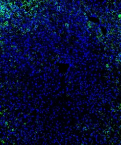
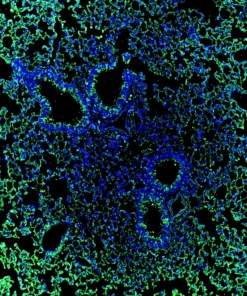
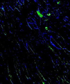
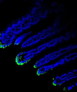
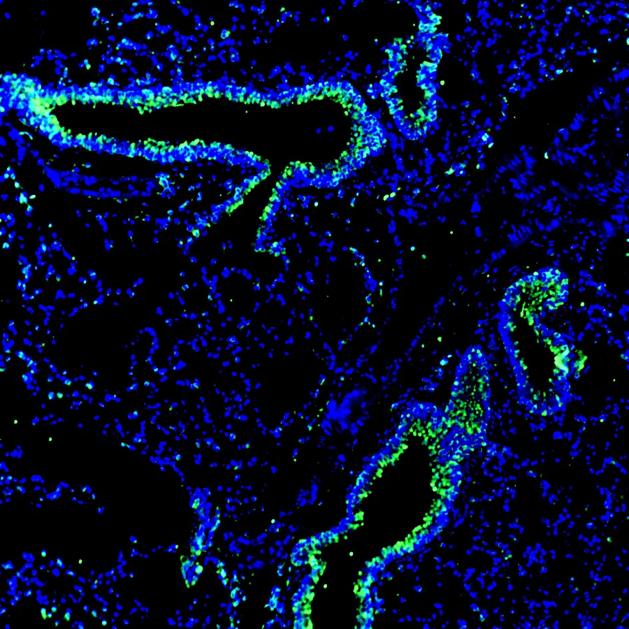
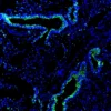
Immunofluorescence staining of mouse lung tissue demonstrated robust endogenous PNR expression within the bronchial epithelial layer. Strong green fluorescence was observed throughout epithelial cells lining the airways, while nuclei were counterstained with DAPI (blue). The staining pattern indicates specific detection of endogenous PNR and supports the suitability of XM0028 for immunofluorescence applications in tissue sections.
| Size | 20 ul, 100 ul |
|---|
Related products